
CMRR
Center for Magnetic Resonance Research, Department of Radiology
News
You are here
SWIFT detection of nanoparticles
SWIFT on the cover of this month's Magnetic Resonance in Medicine.
June 1, 2015 SWIFT detection of nanoparticles makes the cover of this month's Magnetic Resonance in Medicine.
Purpose
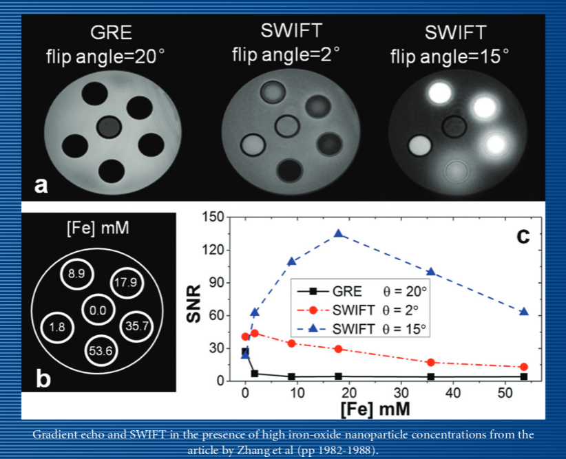
Iron-oxide nanoparticles (IONPs) have proven utility as contrast agents in many MRI applications. Previous quantitative IONP mapping has been performed using mainly T2* mapping methods. However, in applications requiring high IONP concentrations, such as magnetic nanoparticles based thermal therapies, conventional pulse sequences are unable to map T2* because the signal decays too rapidly. In this article, sweep imaging with Fourier transformation (SWIFT) sequence is combined with the Look-Locker method to map T1 of IONPs in high concentrations.
Methods
T1 values of agar containing IONPs in different concentrations were measured with the SWIFT Look-Locker method and with inversion recovery spectroscopy. Precisions of Look-Locker and variable flip angle (VFA) methods were compared in simulations.
Results
The measured R1 (=1/T1) has a linear relationship with IONP concentration up to 53.6 mM of Fe. This concentration exceeds concentrations measured in previous work by almost an order of magnitude. Simulations show SWIFT Look-Locker method is also much less sensitive to B1 inhomogeneity than the VFA method.
Conclusion
SWIFT Look-Locker can accurately measure T1 of IONP concentrations ≤53.6 mM. By mapping T1 as a function of IONP concentration, IONP distribution maps might be used in the future to plan effective magnetic nanoparticle hyperthermia therapy. Magn Reson Med 71:1982–1988, 2014. © 2014 Wiley Periodicals, Inc.