
CMRR
Center for Magnetic Resonance Research, Department of Radiology
Research Highlights - Breast Imaging
You are here
Body Imaging with Spectroscopy: Breast Studies
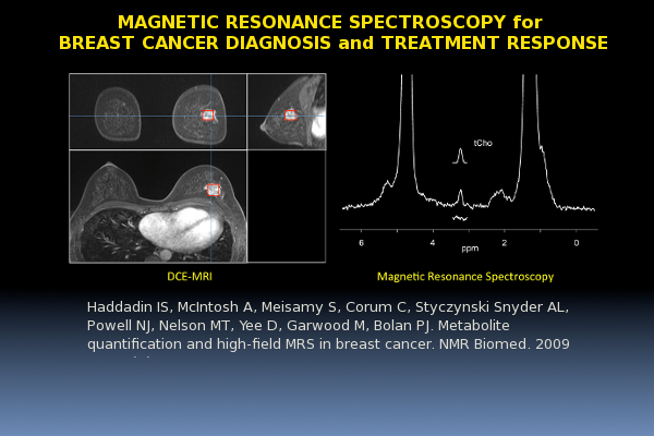
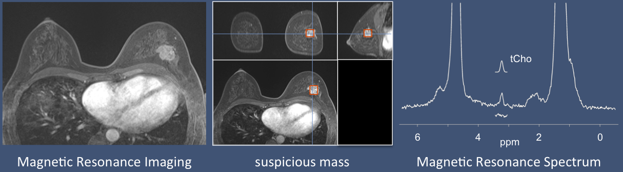
Magnetic Resonance Spectroscopy for Breast Cancer Diagnosis and Treatment Response
Breast cancer is a very common disease, affecting 1 in 8 American women in their lifetimes and causing more than 40,000 deaths each year. Magnetic resonance imaging (MRI) has become the premier imaging method for breast cancer, and is now widely used for diagnosis, staging, assessing treatment response, and screening high-risk patients. Magnetic resonance spectroscopy (MRS) is an additional technique that can be used to improve the accuracy of a breast MRI study. After imaging a breast with conventional MRI methods, a chemical “spectrum” can be measured in a suspicious region, providing additional information about the chemical content of that region. This information has been shown to be helpful in distinguishing benign from malignant masses, and can be used to determine if a mass is responding to chemotherapy treatments faster than MRI alone.
The University of Minnesota’s Center for Magnetic Resonance Research has been instrumental in the development of breast MRS. Lead by Michael Garwood and Patrick Bolan, the CMRR breast team has developed key methodologies that have lead the standardization of breast MRS, including novel acquisition techniques [1], RF coil designs for high-field scanning [2] artifact suppression methods [3][4], and techniques for accurately quantifying the chemical concentrations in breast cancers [5]. Using these techniques, they have performed clinical studies demonstrating how breast MRS can improve diagnostic accuracy [6] and monitor treatment response [7]. These methods have since been incorporated into an NCI-sponsored multisite clinical trial to determine if the methods can be translated into clinical practice.
References
[1] Garwood M, DelaBarre L. The return of the frequency sweep: designing adiabatic pulses for contemporary NMR. J Magn Reson 2001; 153(2) 155-77
[2] Vaughan JT, Snyder CJ, DelaBarre LJ, Bolan PJ, Tian J, Bolinger L, Adriany G, Anderson P, Strupp J, Ugurbil K. Whole-body Imaging at 7T: Preliminary Results. Magn Reson Med 2009; 61(1): 244-8
[3] Bolan PJ, DelaBarre L, Baker EH, Merkle H, Everson LI, Yee D, Garwood M. Eliminating Spurious Sidebands in 1H MRS of Breast Lesions. Magn Reson Med 2002; 48:215-222
[4] Bolan PJ, Henry PG, Baker EH, Meisamy S, Garwood M. Measurement and correction of respiration-induced B0 variations in breast 1H MRS at 4 Tesla. Magn Reson Med 2004; 52:1239-1245
[5] Bolan PJ, Meisamy S, Baker EH, Lin J, Emory T, Nelson M, Everson LI, Yee D, Garwood M. In vivo quantification of choline compounds in the breast with 1H MR spectroscopy. Magn Reson Med 2003; 50:1134-1143
[6] Meisamy S, Bolan PJ, Baker EH, Pollema MG, Le CT, Kelcz F, Lechner MC, Luikens BA, Carlson RA, Brandt KR, Amrami KK, Nelson MT, Everson LI, Emory TH, Tuttle TM, Yee D, Garwood M. Adding in Vivo Quantitative 1H MR Spectroscopy to Improve Diagnostic Accuracy of Breast MR Imaging: Preliminary Results of Observer Performance Study at 4.0 T. Radiology 2005; 236(2):465-475
[7] Meisamy S, Bolan PJ, Baker EH, Bliss RL, Gulbahce E, Everson LI, Nelson MT, Emory TH, Tuttle TM, Yee D, Garwood M. Neoadjuvant chemotherapy of locally advanced breast cancer: predicting response with in vivo 1H MR spectroscopy--a pilot study at 4 T. Radiology 2004; 233:424-431
Haddadin IS, McIntosh A, Meisamy S, Corum C, Styczynski Snyder AL, Powell NJ, Nelson MT, Yee D, Garwood M, Bolan PJ. Metabolite quantification and high-field MRS in breast cancer. NMR Biomed. 2009 Jan;22(1):65-76.
