
CMRR
Center for Magnetic Resonance Research, Department of Radiology
Research Highlights - Multichannel Brain Imaging
You are here
Although ultrahigh magnetic fields (7 Tesla and greater) provide numerous advantages for MRI, it also poses numerous challenges. This BTRC has been focused on developing new imaging methods and engineering solutions towards overcoming these difficulties.
The advantages of high and ultrahigh field fMRI have largely been exploited in applications where a subsection of the brain, such as the primary visual cortex (e.g. [1-3] or the auditory cortex (e.g. [4]) was studied using field of view restriction. Ultimately, however, we need to cover the entire brain in high resolution using Echo Planar Imaging (EPI) that is the preferred imaging method in functional imaging (fMRI) or imaging of connectivity based on water diffusion (DTI, DSI, HARDI etc.). Only recently, this has been possible at magnetic field of 7 Tesla using highly parallel image acquisition (Figure 1). However, high resolution whole brain coverage even with a rapid imaging method like EPI as shown in Figure 1 still takes too long to cover the whole brain.
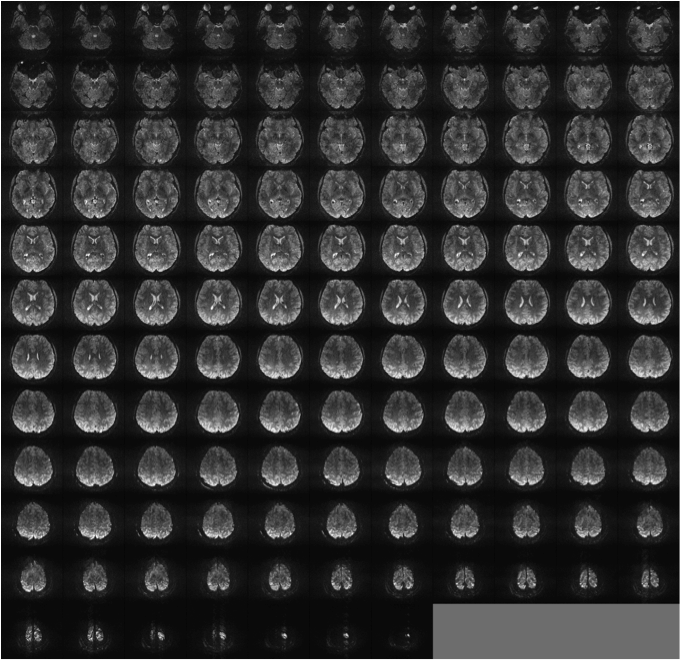
FIGURE 1: RAPID EPI IMAGING of WHOLE BRAIN at 7 TESLA: SINGLE SHOT GE-EPI, 0.75 mm isotropic, Acceleration along Phase encode= 4, partial Fourier=6/8; 256 x 256; 128slices TE=20msec
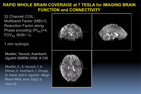
To overcome this, we recently developed, as part of the BTRC supported effort, “slice accelerated” multiband (MB) EPI imaging for fMRI [5, 6]. In this technique, several slices are simultaneously excited using multibanded RF pulses and subsequently acquired in a single EPI echo train; the simultaneously acquired slices are then unaliased using spatial information inherent in signal detection with a multi-coil array.
Simultaneous acquisition of multiple slices using Gradient Recalled Echo (GRE) and subsequently unfolding them using parallel imaging principles was previously described for spine imaging [7] in 2001. This GRE approach was further pursued in the CAIPIRINHA [8, 9] technique where unaliasing of slices were improved by manipulating the phase of the RF excitation pulses progressively for each k-space line in the GRE sequence. Following this work, a method employing gradients “blips” to achieve a similar shift in multiband EPI imaging was also described in an abstract [10].
We have further improved on this approach and developed new image reconstruction strategies to achieve high MB factors in the presence of also high conventional accelerations along the phase encode dimension. The latter is absolutely mandatory to attain high quality EPI images without significant signal loss or distortion (unpublished results).
REFERENCES
1. Yacoub, E., A. Shmuel, N. Logothetis, and K. Ugurbil: Robust detection of ocular dominance columns in humans using Hahn Spin Echo BOLD functional MRI at 7 Tesla. Neuroimage, 2007. 37(4): p. 1161-77.
2. Yacoub, E., N. Harel, and K. Ugurbil: High-field fMRI unveils orientation columns in humans. Proc Natl Acad Sci U S A, 2008. 105(30): p. 10607-12.
3. Olman, C.A., P.F. Van de Moortele, J.F. Schumacher, J.R. Guy, K. Ugurbil, and E. Yacoub: Retinotopic mapping with spin echo BOLD at 7T. Magn Reson Imaging, 2010. 28(9): p. 1258-69.
4. Formisano, E., D.S. Kim, F. Di Salle, P.F. van de Moortele, K. Ugurbil, and R. Goebel: Mirror-symmetric tonotopic maps in human primary auditory cortex. Neuron, 2003. 40(4): p. 859-69.
5. Moeller, S., E. Auerbach, P.-F. van de Moortele, G. Adriany, and K. Ugurbil: fMRI with 16 fold reduction using multibanded multislice sampling. Proc. Int. Soc. Magn. Reson. in Med. , 2008. 16: p. 2366.
6. Moeller, S., E. Yacoub, C.A. Olman, E. Auerbach, J. Strupp, N. Harel, and K. Ugurbil: Multiband multislice GE-EPI at 7 tesla, with 16-fold acceleration using partial parallel imaging with application to high spatial and temporal whole-brain fMRI. Magn Reson Med, 2010. 63(5): p. 1144-53.
7. Larkman, D.J., J.V. Hajnal, A.H. Herlihy, G.A. Coutts, I.R. Young, and G. Ehnholm: Use of multicoil arrays for separation of signal from multiple slices simultaneously excited. J Magn Reson Imaging, 2001. 13(2): p. 313-7.
8. Breuer, F.A., M. Blaimer, R.M. Heidemann, M.F. Mueller, M.A. Griswold, and P.M. Jakob: Controlled aliasing in parallel imaging results in higher acceleration (CAIPIRINHA) for multi-slice imaging. Magnetic resonance in medicine : official journal of the Society of Magnetic Resonance in Medicine / Society of Magnetic Resonance in Medicine, 2005. 53(3): p. 684-91.
9. Breuer, F.A., M. Blaimer, M.F. Mueller, N. Seiberlich, R.M. Heidemann, M.A. Griswold, and P.M. Jakob: Controlled aliasing in volumetric parallel imaging (2D CAIPIRINHA). Magnetic resonance in medicine : official journal of the Society of Magnetic Resonance in Medicine / Society of Magnetic Resonance in Medicine, 2006. 55(3): p. 549-56.
10. Nunes, R.G., J.V. Hajnal, X. Golay, and D.J. Larkman: Simultaneous slice excitation and reconstruction for single shot EPI. Proc. Int. Soc. Mag Reson Med, 2006. 14: p. 293.
