
CMRR
Center for Magnetic Resonance Research, Department of Radiology
Research Highlights - Visual Cortex Vasculature
You are here
Vascular Imaging at Ultra-high Fields
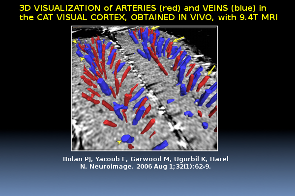
An example of a 3D surface rendering of the microvasculature vessels in the cat visual cortex, obtained in-vivo, on a 9.4T MRI machine. The spatial distribution of the arteries and veins indicated by red and blue arrows (respectively). (Bolan et al., NeuroImage 2006)
Building on the advantages of high-field MRI, it is feasible to image and classify cortical vasculature, in vivo, at resolutions approaching the microscopic scale. Maximum Intensity Projections (MIPs) of a 3D volume of a cat cortical vasculature is shown in axial orientations. The image clearly shows the organization of intracortical vessels. (Bolan et al.,NeuroImage 2006)
REFERENCES
In vivo micro-MRI of intracortical neurovasculature. Bolan PJ, Yacoub E, Garwood M, Ugurbil K, Harel N. Neuroimage. 2006 Aug 1;32(1):62-9.
