CMRR
Center for Magnetic Resonance Research, Department of Radiology
Faculty and Staff
You are here
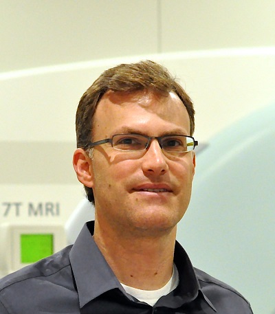
Dr. Gregory J. Metzger (Greg Metzger)
|
|
|
Dr. Metzger received his B.S. from the University of Pennsylvania in 1992 and his Ph.D. from the Department of Biomedical Engineering at the University of Minnesota in 1997. His graduate research, conducted under the mentorship of Dr. Xiaoping Hu, focused on novel magnetic resonance chemical shift imaging techniques. After his graduate studies, Dr. Metzger accepted a position with Philips Medical Systems as a clinical scientist at the University of Texas Southwestern Medical Center in Dallas. In this position, he expanded his knowledge in clinical research with specific work on breast, kidney, cardiac and brain MRI applications. In his final two years of his eight year tenure with Philips, he worked at NIH as a senior clinical scientist focusing on diagnostic prostate imaging and MRI guided prostate interventions. With a desire to return to academia, he accepted his current position at the University of Minnesota in 2005 as an associate professor with a joint appointment in the departments of Radiology and Urologic Surgery.
Research Interests:
- Quantitative Prostate Cancer MRI
- Development of Correlative Pathology for predictive model development
- Development of hardware and methods for ultrahigh field Body MRI
My expertise or research interests fall in two sometimes overlapping areas: 1) the development of multi-parametric MRI for cancer characterization and 2) the development of imaging strategies to safely realize the advantages of ultrahigh field MRI for studies in the human torso.
Related to the first area of research, I am investigating the potential of quantitative magnetic resonance imaging and spectroscopy to non-invasively determine the extent and aggressiveness of prostate cancer in clinical studies. Initial research objectives involve the development of a multi-parametric statistical model to non-invasively determine cancer probability maps by using quantitative anatomic and molecular pathology as a gold standard.
My second major area of research involves the development of hardware, RF management strategies and acquisition methods necessary to make imaging at ultra-high magnetic fields feasible in the human torso. While one ultrahigh field imaging goal is the improved characterization of prostate cancer, other areas of investigation have involved the development of dynamic RF shimming strategies for managing non-uniform transmit B1 fields enabling a wide range of studies including non-contrast enhanced perfusion and angiography studies in the prostate, kidneys and heart.
My areas of research require a highly collaborative approach involving clinicians, basic researchers and engineers. This environment as allowed me to mentor several individuals where engineering approaches are used to eventually solve clinical problems. I have taken the lead at our imaging center to promote research opportunities for students from departments outside of Radiology including those from the Medical School and the College of Science and Engineering.
Selected Publications:
Metzger, G. J., Kalavagunta, C., Spilseth, B., Bolan, P. J., Li, X., Hutter, D., Nam, J. W., Johnson, A. D., Henricksen, J. C., Moench, L., Konety, B., Warlick, C. A., Schmechel, S. C. & Koopmeiners, J. S. Development of Quantitative Multiparametric MRI Models for Prostate Cancer Detection using Registered Correlative Histopathology (In Press.)
Wasserman, N. F., Spilseth, B., Golzarian, J. & Metzger, G. J. Use of MRI for Lobar Classification of Benign Prostatic Hyperplasia: Potential Phenotypic Biomarkers for Research on Treatment Strategies. (2015) AJR Am J Roentgenol 205, 564-571.
Li, X., Wang, D., Auerbach, E. J., Moeller, S., Ugurbil, K. & Metzger, G. J. Theoretical and experimental evaluation of multi-band EPI for high-resolution whole brain pCASL Imaging. (2015) NeuroImage 106, 170-181.
Li, X., Bolan, P. J., Ugurbil, K. & Metzger, G. J. Measuring renal tissue relaxation times at 7 T. (2015) NMR Biomed 28, 63-69.
Kalavagunta, C., Zhou, X., Schmechel, S. C. & Metzger, G. J. Registration of in vivo prostate MRI and pseudo-whole mount histology using Local Affine Transformations guided by Internal Structures (LATIS). (2015) J Magn Reson Imaging 41, 1104-1114.
Erturk, M. A., Tian, J., Van de Moortele, P. F., Adriany, G. & Metzger, G. J. Development and evaluation of a multichannel endorectal RF coil for prostate MRI at 7T in combination with an external surface array. (2015) J Magn Reson Imaging Published on line before Print; DOI: 10.1002/jmri.25099.
Rizzardi, A. E., Rosener, N. K., Koopmeiners, J. S., Isaksson Vogel, R., Metzger, G. J., Forster, C. L., Marston, L. O., Tiffany, J. R., McCarthy, J. B., Turley, E. A., Warlick, C. A., Henriksen, J. C. & Schmechel, S. C. Evaluation of protein biomarkers of prostate cancer aggressiveness. (2014) BMC Cancer 14, 244.
Kalavagunta, C., Michaeli, S. & Metzger, G. J. In vitro Gd-DTPA relaxometry studies in oxygenated venous human blood and aqueous solution at 3 and 7 T. (2014) Contrast media & molecular imaging 9, 169-176.
Iltis, I., Choi, J., Vollmers, M., Shenoi, M., Bischof, J. & Metzger, G. J. In vivo detection of the effects of preconditioning on LNCaP tumors by a TNF-alpha nanoparticle construct using MRI. (2014) NMR Biomed 27, 1063-1069.
Metzger, G. J., Auerbach, E. J., Akgun, C., Simonson, J., Bi, X., Ugurbil, K. & van de Moortele, P. F. Dynamically applied B1+ shimming solutions for non-contrast enhanced renal angiography at 7.0 Tesla. (2013) Magn Reson Med 69, 114-126.
Li, X. & Metzger, G. J. Feasibility of measuring prostate perfusion with arterial spin labeling. (2013) NMR Biomed 26, 51-57.
Snyder, C. J., Delabarre, L., Moeller, S., Tian, J., Akgun, C., Van de Moortele, P. F., Bolan, P. J., Ugurbil, K., Vaughan, J. T. & Metzger, G. J. Comparison between eight- and sixteen-channel TEM transceive arrays for body imaging at 7 T. (2012) Magn Reson Med 67, 954-964.
Rizzardi, A. E., Johnson, A. T., Vogel, R. I., Pambuccian, S. E., Henriksen, J., Skubitz, A. P., Metzger, G. J. & Schmechel, S. C. Quantitative comparison of immunohistochemical staining measured by digital image analysis versus pathologist visual scoring. (2012) Diagn Pathol 7, 42.
Metzger, G. J., Dankbar, S. C., Henriksen, J., Rizzardi, A. E., Rosener, N. K. & Schmechel, S. C. Development of multigene expression signature maps at the protein level from digitized immunohistochemistry slides. (2012) Plos ONE 7, e33520.
Metzger, G. J., van de Moortele, P. F., Akgun, C., Snyder, C. J., Moeller, S., Strupp, J., Andersen, P., Shrivastava, D., Vaughan, T., Ugurbil, K. & Adriany, G. Performance of external and internal coil configurations for prostate investigations at 7 T. (2010) Magn Reson Med 64, 1625-1639.
Snyder, C. J., DelaBarre, L., Metzger, G. J., van de Moortele, P. F., Akgun, C., Ugurbil, K. & Vaughan, J. T. Initial results of cardiac imaging at 7 Tesla. (2009) Magn Reson Med 61, 517-524.
Metzger, G. J., Snyder, C., Akgun, C., Vaughan, T., Ugurbil, K. & Van de Moortele, P. F. Local B1+ shimming for prostate imaging with transceiver arrays at 7T based on subject-dependent transmit phase measurements. (2008) Magn Reson Med 59, 396-409.
Ocak, I., Bernardo, M., Metzger, G., Barrett, T., Pinto, P., Albert, P. S. & Choyke, P. L. Dynamic contrast-enhanced MRI of prostate cancer at 3 T: a study of pharmacokinetic parameters. (2007) AJR Am J Roentgenol 189, 849.
Complete List of Published Work in My NCBI:

