
CMRR
Center for Magnetic Resonance Research, Department of Radiology
Research Highlights - Cartilage Assessment at 3T
You are here
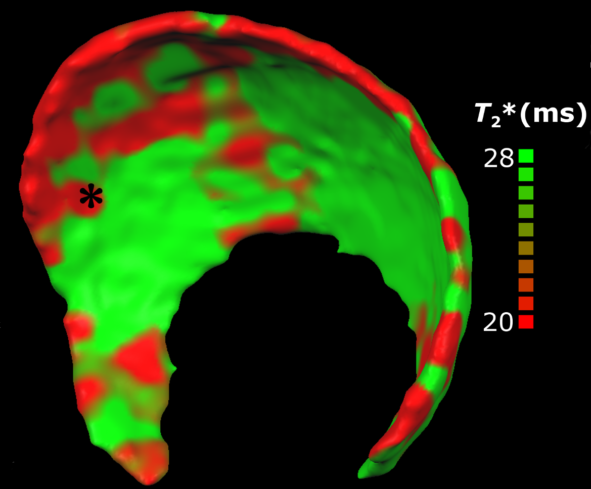
HIP MRI at 3 Tesla T2* for patient specific cartilage assessment in Femoroacctabular Impingment (FAI).
Representative GRE – Images (TE 11 ms), which is used in conjunction with 5 other echo times to calculate the T2 * map. Note the high resolution with clear separation of the superolateral acetabular cartilage from the articular cartilage of the femoral head.
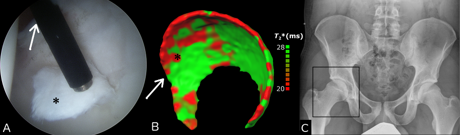
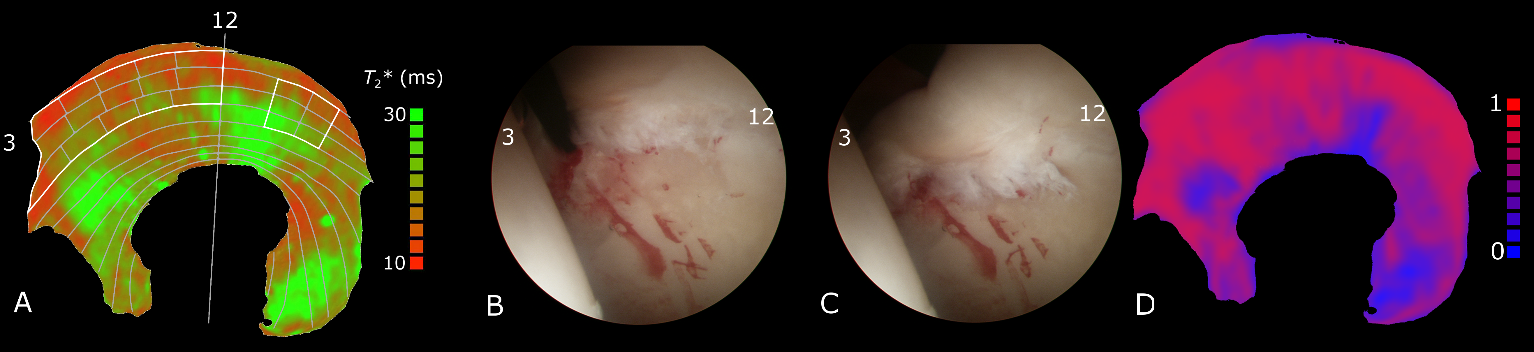
3T Results - T2* mapping and arthroscopic correlation.
