
CMRR
Center for Magnetic Resonance Research, Department of Radiology
Research Highlights - Imaging Orientation Domains in the Primary Visual Cortex
You are here


  |
Functional Brain Imaging (fMRI) was introduced by concurrent and independent work in two laboratories [1, 2], one of which is this BTRC. Since its introduction, fMRI has fueled explosive developments in our understanding of brain function in health and disease. fMRI rapidly emerged as the dominant and virtually indispensible method for studying the human brain, which is endowed with unique capabilities that often cannot be studied in animal models. It has been possible to visualize human brain activity in three dimensions, in the whole brain with approximately a millimeter resolution or in subsections of the brain in with submillimeter resolution; an example of the latter coming from this BTRC is the imaging of orientation columns that were never previously visualized in the human brain.
Orientation columns but were successfully mapped with fMRI at 7T [3] in the primary visual area V1 together with ocular dominance columns (ODC) revealing the organizational relationship between them [3] (Figure 1).
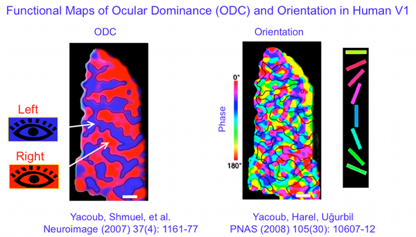
FIGURE 1: Human Ocular Dominance Columns (ODCs) and Orientation Columns imaged in the same subject.
While a lot has been known about human ODCs for quite some time due to the ability to image them even in the postmortem brain, orientation columns require functional methods for their depiction and were never imaged in the human brain before. This study heralds the exciting prospect of exploring unmapped and/or unknown functional organizations in the human brain at the level of elementary computational units. This has been possible only with ultrahigh magnetic fields, the central technological pursuit in the BTRC located in CMRR, and extensive theoretical and experimental studies aimed at understanding the complex set of mechanisms underlying the ultimate coupling between neuronal activity and MRI signals.
REFERENCES
1. Kwong, K.K., J.W. Belliveau, D.A. Chesler, I.E. Goldberg, R.M. Weisskoff, B.P. Poncelet, D.N. Kennedy, B.E. Hoppel, M.S. Cohen, R. Turner, and et al.: Dynamic magnetic resonance imaging of human brain activity during primary sensory stimulation. Proc Natl Acad Sci U S A, 1992. 89(12): p. 5675-9.
2. Ogawa, S., D.W. Tank, R. Menon, J.M. Ellermann, S.G. Kim, H. Merkle, and K. Ugurbil: Intrinsic signal changes accompanying sensory stimulation: functional brain mapping with magnetic resonance imaging. Proc Natl Acad Sci U S A, 1992. 89(13): p. 5951-5.
3. Yacoub, E., N. Harel, and K. Ugurbil: High-field fMRI unveils orientation columns in humans. Proc Natl Acad Sci U S A, 2008. 105(30): p. 10607-12.
