
CMRR
Center for Magnetic Resonance Research, Department of Radiology
Neuromodulation
You are here
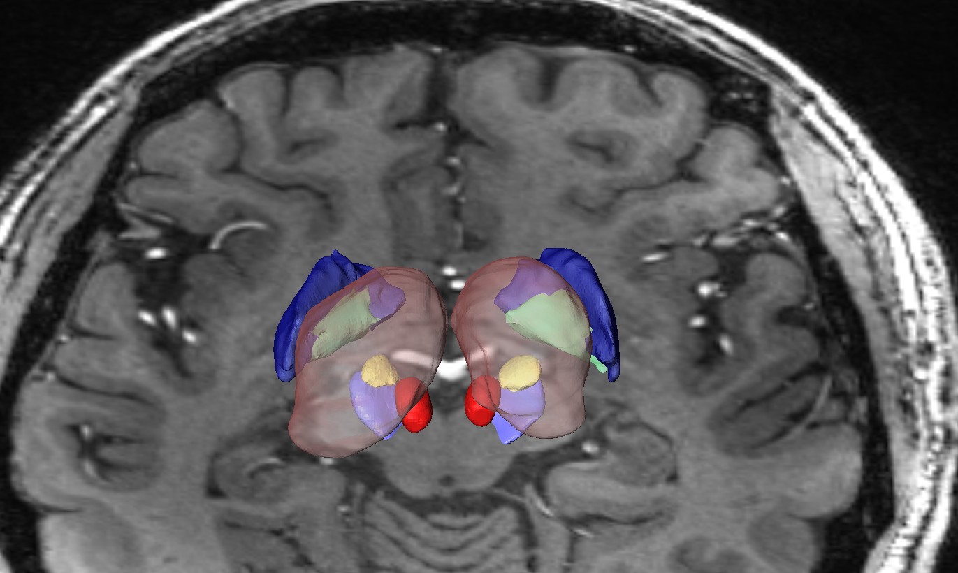
Deep Brain Stimulation:
Using a 7 Tesla MRI, a 3D volume renderings of the patient’s own brain anatomy is generated. Nuclei of the Basal Ganglia are shown: globus pallidus (PGi: green; GPe: Blue), red nucleus (red), subthalamic nucleus (yellow) and substantia nigra (light blue).
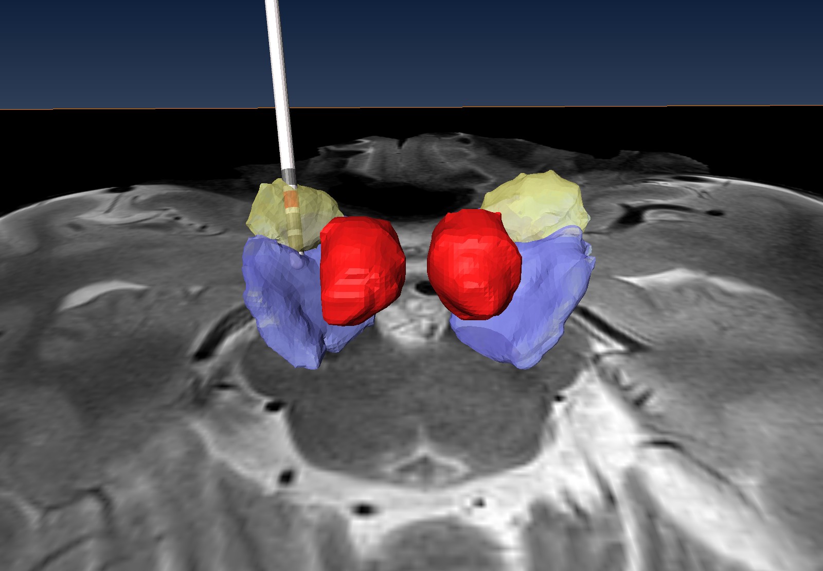
STN-DBS targeting:
An anatomical model of a Parkinson patient after DBS surgery. The DBS lead (white) is fused with an MR image acquired at 7T showing the electrode location within the STN. Red nucleus (red), subthalamic nucleus (STN, yellow) and substantia nigra (light blue).
