
CMRR
Center for Magnetic Resonance Research, Department of Radiology
Research Highlights
You are here
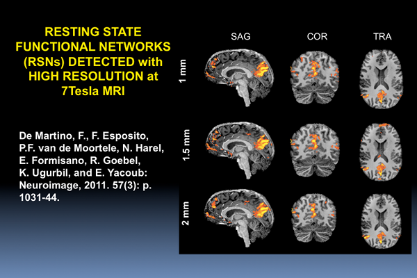
We have recently demonstrated high resolution, whole brain analysis of resting state networks (RSNs) in the human brain using ultrahigh field (7 Tesla) functional imaging [1]. Study of RSNs is important in studies of functional connectivity in the brain under normal and pathological conditions.
An fMRI time series collected in the absence of tasking or stimulation, i.e. with the subject simply at “rest” in the scanner, reveals low-frequency (<0.1 Hz) fluctuations in the brain that often display strong correlations among distant regions (e.g. [2-5]), revealing networks of correlated variations referred to as resting state networks (RSNs);
Detailed RSNs has been restricted primarily to 3 Tesla or lower magnetic fields using low resolution (voxel volumes of approximately 27 mm3, i.e. 3 mm isotropic voxels) to achieve sufficient minimum signal-to-noise and functional contrast-to-noise ratio (SNR and fCNR, respectively). Furthermore, typically, these studies employ spatial smoothing to reduce noise, and often also resort to group averaging, which further amplifies spatial blurring by requiring additional smoothing.
The higher functional specificity, and fCNR inherent at ultrahigh fields such as 7 Tesla can be exploited to alleviate this limitation. This was accomplished recently, demonstrating detailed RSNs with 1, 1.5. and 2 mm isotropic spatial resolution in single subjects without spatial smoothing and in group averages. By comparing results between multiple resolutions we show that the smaller voxels volumes (1 and 1.5 mm isotropic) result in reduced partial volume effects, permitting separations of detailed spatial features within the default mode network (DMN) patterns as well as a better function to anatomy correspondence. demonstrating advantages.
REFERENCES
1. De Martino, F., F. Esposito, P.F. van de Moortele, N. Harel, E. Formisano, R. Goebel, K. Ugurbil, and E. Yacoub: Whole brain high-resolution functional imaging at ultra high magnetic fields: An application to the analysis of resting state networks. Neuroimage, 2011. 57(3): p. 1031-44.
2. Biswal, B., F.Z. Yetkin, V.M. Haughton, and J.S. Hyde: Functional connectivity in the motor cortex of resting human brain using echo-planar MRI. Magnetic Resonance in Medicine, 1995. 34(4): p. 537-41.
3. Lowe, M.J., B.J. Mock, and J.A. Sorenson: Functional connectivity in single and multislice echoplanar imaging using resting-state fluctuations. Neuroimage, 1998. 7(2): p. 119-32.
4. Damoiseaux, J.S., S.A. Rombouts, F. Barkhof, P. Scheltens, C.J. Stam, S.M. Smith, and C.F. Beckmann: Consistent resting-state networks across healthy subjects. Proceedings of the National Academy of Sciences of the United States of America, 2006. 103(37): p. 13848-53.
5. Nir, Y., U. Hasson, I. Levy, Y. Yeshurun, and R. Malach: Widespread functional connectivity and fMRI fluctuations in human visual cortex in the absence of visual stimulation. Neuroimage, 2006. 30(4): p. 1313-24.
6. Yacoub, E., T.Q. Duong, P.F. Van De Moortele, M. Lindquist, G. Adriany, S.G. Kim, K. Ugurbil, and X. Hu: Spin-echo fMRI in humans using high spatial resolutions and high magnetic fields. Magn Reson Med, 2003. 49(4): p. 655-64.