CMRR
Center for Magnetic Resonance Research, Department of Radiology
Research Highlights - Novel MRI: SWIFT
You are here
SWIFT MRI: Imaging Hard and Soft Dental Tissues
Seeing the Invisible
|
The goal of TRD 3 is to expand significantly the capabilities of MRI by exploiting the unique features of frequency-modulated (FM) pulses. Our new technique called SWIFT (SWeep Imaging with Fourier Transformation) is a radically different approach to MRI with many unique and powerful features, including nearly silent operation and the ability to visualize hard tissues (e.g., cortical bone, enamel, dentin) [1]. These capabilities are expanding the utility of MRI into areas of research and clinical application where previously it played little or no role. |
 |
How SWIFT works:
The major breakthrough underlying SWIFT comes from the use of time-shared excitation and acquisition to permit capture of signals originating from molecules with extremely short transverse relaxation times. For example, water molecules in teeth have T2 values in the range of only 50 to 500 microseconds, whereas most soft tissues have T2 values in the range of tens of milliseconds. The idea for gapped excitation and detection to capture ultrafast relaxing MRI signals originated in TRD 3, but required solving the technical challenges of gapped acquisition, which was accomplished.
History of SWIFT
In 2002, Dr. Garwood had the idea that it might be possible to acquire spatially-encoded MR signals while simultaneously applying a frequency-modulated (FM) pulse. He tested the idea using Bloch simulations, and later the same year, experimentally demonstrated it by acquiring steady-state MR signals during gaps inserted into FM pulses. These preliminary data were included in the competitive renewal of the NIH BTRC grant that was submitted in December, 2002 and funded in 2003.
An Example of SWIFT's Unique Capabilities: Dental MRI
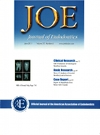
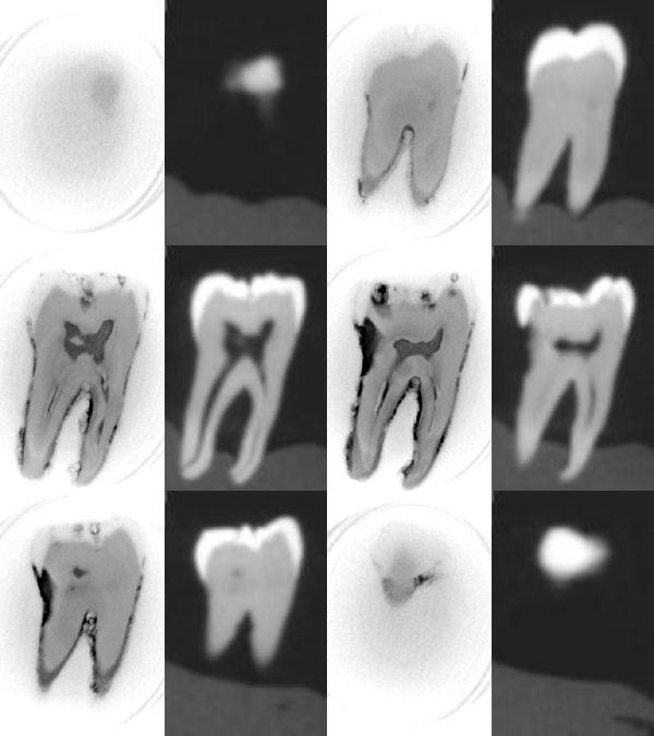
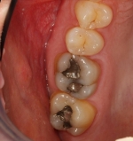
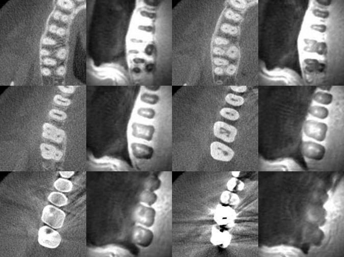
Our recent publication in Journal of Endodontics (cover article) demonstrates the concept that SWIFT has significant diagnostic potential for dentistry [2]. SWIFT has potential to address many long-standing imaging issues of great consequence to dentistry. First and foremost is the simultaneous (in the same scan) imaging of hard and soft dental tissues, both of which are important for diagnostic purposes. Beyond carious lesion detection, SWIFT has the ability to simultaneously assess pulpal and periodontal tissues, which are other areas with significant long-standing diagnostic challenges. We have imaged in vitro teeth at high-resolution, which allows for the identification of minute anatomical and pathological features (Fig 1), and we have produced low-resolution in vivo images from a normal volunteer (Fig 2).
SWIFT versus cone-beam CT |
|||
 |
|||
| Inverted | |||
| SWIFT 0.1 mm | CBCT 0.2 mm | ||
| Fig 1 | |||
In vivo teeth imaging |
||
 |
 |
|
| metallic restorations (amalgam) |
CBCT | SWIFT 10 min |
| Fig 2 | ||
| Idiyatullin et al, J Endod, 2011 (cover article) | ||
Significance:
Dental imaging techniques are routinely used in all aspects of patient care, from diagnosis to risk assessment, as well as pre-prosthetic planning and monitoring treatment outcomes [3]. Detection and diagnosis of caries is significant for the U.S. population because caries is the most common chronic childhood disease, being five times more common than asthma [4]. Currently, there is no single diagnostic imaging modality in clinical use that has the potential to address the issues of early detection (screening), three-dimensional determination of the extent of carious lesions (staging of disease), and measurement of pulpal involvement (prognostic measure of treatment outcome), which are the three major applications of diagnostic imaging in healthcare [5].
LITERATURE CITED
[1] Idiyatullin D, Corum C, Park JY, Garwood M. Fast and quiet MRI using a swept radiofrequency. J Magn Reson 2006:181(2):342-9 Epub 2006 Jun 19.
[2] Idiyatullin D, Corum C, Moeller S, Prasad H, Garwood M, Nixdorf D. Dental Magnetic Resonance Imaging: Making the Invisible Visible. J Endod 2011:37:745-52.
[3] White SC, Pharoah MJ. Principles of radiographic interpretation. Oral Radiology: Principles and Interpretation. St. Louis, MO: Mosby Elsevier, 2009:256-69.
[4] U.S. Department of Health and Human Services. Oral Health in America: A Report of the Surgeon General. Rockville, MD: U.S. Department of Health and Human Services, National Institute of Dental and Craniofacial Research, National Institutes of Health, 2000.
[5] Hillman BJ, Gatsonis CA. When is the right time to conduct a clinical trial of a diagnostic imaging technology? Radiology 2008:248(1):12-5.
Dental MRI: Making the Invisible Visible. D. Idiyatullin, C. Corum, S. Moller, H.S. Prasad, M. Garwood, and D.R. Nixdorf, J. Endod 37:745-752, 2011. (Cover Article)