
CMRR
Center for Magnetic Resonance Research, Department of Radiology
Research Highlights
You are here

Focal hippocampal dysfunction initiates electrophysiological seizures and impairs cognition in mesial temporal lobe epilepsy (TLE). The lesion most commonly reported in surgical and autopsy series of mesial TLE is hippocampal sclerosis (HS).
Hippocampal pathology in HS is normally studied using tissue examination of surgical specimens and during autopsies. Such studies reveal that HS affects subregions of the hippocampus differentially: Ammon’s horn sclerosis (loss of pyramidal neurons predominantly in the CA1 region) is more commonly encountered than is end-folium sclerosis (neuronal loss in CA4 and the adjacent hilus of the dentate gyrus), and they co-exist in severe TLE, as “total” HS.
An in vivo technique that defined subregional distributions of hippocampal atrophy and other fine detail of the hippocampus could be used to to better select TLE patients who should (or should not) have epilepsy surgery.
Cerebral MRI at 1.5T and 3T does not reliably image the major intrahippocampal structures, because of both submillimetric dimensions and limited MR contrast in these tissues. Ultrahigh field (7T and higher) MRI offers greatly increased signal-to-noise, however, allowing for increased spatial resolution and increased tissue contrast compared with 1.5 and 3 T fields.
Figure below shows 7T T2-weighted MRI, coronal slice cutting through the hippocampal head region, for a control subject (c6) and three patients.
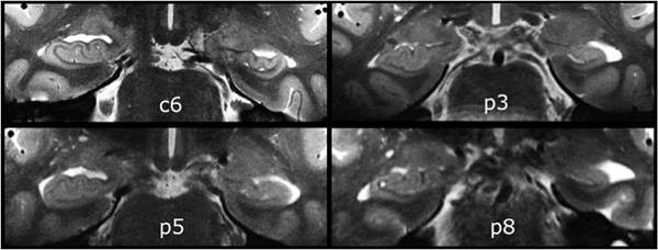
In this 7T study, we demonstrate that
1. Intrahippocampal structures are consistently detected in healthy subjects and those with HS, permitting subregional hippocampal volumetry, with 7 T MRI.
2. In sclerotic hippocampi, 7T MRI can distinguish predominantly Ammon’s horn atrophy from atrophy that also involves the dentate gyrus.
3. Deformity of the hippocampal head featuring paucity of digitations can be detected in the presence or absence of hippocampal atrophy, using 7 T MRI.
4. The internal structures of malrotated hippocampal bodies were clearly visible, using 7T MRI.
These findings indicate that the increases in spatial resolution and contrast of 7 T MRI should significantly increase the sensitivity and specificity of structural imaging in presurgical evaluation of medically refractory TLE.