CMRR
Center for Magnetic Resonance Research, Department of Radiology
Research Highlights
You are here
A novel imaging technique, RASER, for fMRI

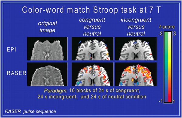
In functional MRI (fMRI), tiny signal changes in response to neuronal activity are detected in the brain. In certain brain regions, such as, the orbitofrontal cortex (OFC), which is located above the eyes behind the forehead, the conventional imaging method for fMRI referred to as EPI shows severe image distortions and signal loss as result of local magnetic field variation near the air-tissue interface of the sinus (top row, left image – axial slice through the OFC of a human subject at 7 T). As a consequence, functional studies of this brain regions, which are involved in decision-making and planning, are technically very challenging especially at ultrahigh magnetic fields. The two activation maps in the top row show the failure of EPI to detect neuronal activity in the OFC of a human subject, who performs color-word match Stroop task engaging the OFC. In novel imaging technique, RASER (rapid acquisition by sequential excitation and refocusing) for ultrafast imaging has been developed at CMRR. This imaging approach is inherently insensitive to magnetic field variations, which cause severe image artifacts in EPI (bottom row left image – same slice location as in top row). RASER also shows superior detection sensitivity of neuronal activity compared to EPI in the same subject performing the same Stroop-task (bottom row – two right activation maps).