
CMRR
Center for Magnetic Resonance Research, Department of Radiology
Research Highlights
You are here
Engineering Core Highlights
The Engineering Core specializes in developing and supporting new technology for the many biomedical applications of high field MRI at the CMRR. For a significant collection of the world’s highest field magnets (Figure 1), the engineers must develop and design new head coils (Figure 3), body coils (Figure 4) and assure their safe and effective use (Figure 2).
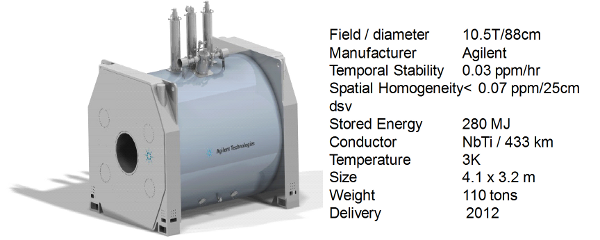
Figure 1. 10.5T Whole body human magnet to be delivered in 2012
Engineering design begins with modeling. In Figure 2a, electromagnetic propagation and loss is numerically calculated for the B1 field, E field, specific absorption rate (SAR), heating rate dT, and temperature T, for the whole human body exposed to the ultra-high frequencies required to image the body at ultra-high field strengths. (1)
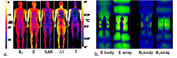
Figure 2. RF propagation and loss calculations for ultra-high field MRI.
Figure 2b shows the graphical electrodynamics of ultra-high-frequency excitation of the body loaded magnet bore space inside a whole body coil, and with a local array. These unique models are critical to understanding and designing both hardware and experimental protocols for safe and successful imaging and spectroscopy at unprecedented field strengths.
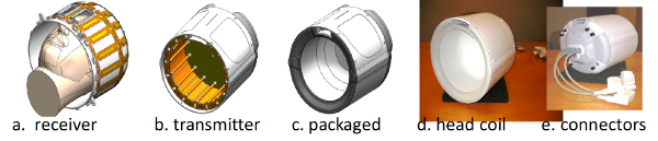
Figure 3. RF head coil components for ultra-high field MRI.
To compensate for shortwave losses and interference patterns in the anatomy, and to enhance radiofrequency (RF) localization and parallel imaging performance, engineers and physicists at the CMRR have invented and employed techniques such as B1 shimming (2) (3) (4), and RF frontend systems incorporating multi-channel transmission. (5) (6) The head coils of Figure 3 and the body coils of Figure 4 demonstrate these advances. In Figure 3, a multi-channel receiver “a” fits into a multi-channel transmitter “b” and is packaged as “c”. The real coil “d” is connectorized for MRI system interface “e”. (7) (8)
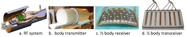
Figure 4. Body coil components for ultra-high field MRI.
Ultra-high field whole body imaging has also been pioneered at the CMRR. New methods and technology were developed to make this possible. The conceptual drawing of “a” shows a three dimensional, multi-channel body coil used for transmit realized in “b,” enclosing a multi-channel receiver array demonstrated in “c”. A more efficient alternative, the transceiver array is shown in Figure 4d. Typically a pair of receivers “c” or transceivers “d” are used in anterior and posterior positions for body imaging. (9) (10) (11) (12)
REFERENCES
1. Vaughan JT, Snyder CJ, DelaBarre LJ, Bolan PJ, Tian J, Bolinger L, Adriany G, Andersen P, Strupp J, Ugurbil K. Whole-body imaging at 7T: Preliminary results. Magn Reson Med 2009;61(1):244-248.
2. Van de Moortele PF, Akgun C, Adriany G, Moeller S, Ritter J, Collins CM, Smith MB, Vaughan JT, Ugurbil K. B1 destructive interferences and spatial phase patterns at 7 T with a head transceiver array coil. Magn Reson Med 2005;54(6):1503-1518.
3. Vaughan JT, Adriany G, Snyder C, Tian J, Thiel T, Bolinger L, Liu H, DelaBarre L, Ugurbil K. Efficient high-frequency body coil for high-field MRI. Magn Reson Med 2004;52:851-859.
4. Vaughan JT, Hetherington H, Otu J, Pan J, Pohost G. High frequency volume coils for clinical nuclear magnetic resonance imaging and spectroscopy. Magn Reson Med 1994;32:206-218.
5. Vaughan T, DelaBarre L, Snyder C, Tian J, Akgun C, Shrivastava D, Liu W, Olson C, Adriany G, Strupp J, Andersen P, Gopinath A, van de Moortele PF, Garwood M, Ugurbil K. 9.4T Human MRI: Preliminary Results. Magn Reson Med 2006;56(6):1274-1282.
6. Adriany G, Van de Mortele P-F, Wiesinger F, Moeller S, Strupp J, Andersen P, Snyder C, Zhang X, Chen W, Pruessmann K, Boesiger P, Vaughan J, Ugurbil K. Transmit and receive transmission line arrays for 7 tesla parallel imaging. Magn Reson Med 2005;53(2):434-445.
7. Vaughan JT, Garwood M, Collins CM, Liu W, DelaBarre L, Adriany G, Andersen P, Merkle H, Goebel R, Smith MB, Ugurbil K. 7T vs. 4T: RF power, homogeneity, and signal-to-noise comparison in head images. Magn Reson Med 2001;46(1):24-30.
8. Adriany G, Auerbach EJ, Snyder CJ, Gozubuyuk A, Moeller S, Ritter J, Van de Moortele PF, Vaughan T, Ugurbil K. A 32-Channel Lattice Transmission Line Array for Parallel Transmit and Receive MRI at 7 Tesla. Magn Reson Med 2010;63(6):1478-1485.
9. Snyder CJ, DelaBarre L, Metzger GJ, van de Moortele PF, Akgun C, Ugurbil K, Vaughan JT. Initial results of cardiac imaging at 7 Tesla. Magn Reson Med 2009;61(3):517-524.
10. Snyder C, DelaBarre L, Moeller S, Akgun C, Van de Moortele P-F, Bolan P, Ugurbil K, Vaughan JT, Metzger G. Comparison Between Eight- and Sixteen-Channel TEM Transceive Arrays for Body Imaging at 7 Tesla. Magn Reson Med 2011;in press.
11. Suttie JJ, DelaBarre L, Pitcher A, van de Moortele P-F, Dass S, Snyder CJ, Francis JM, Metzger GJ, Weale P, Ugurbil K, Neubauer S, Robson M, Vaughan JT. 7 Tesla (T) human cardiovascular magnetic resonance imaging using FLASH and SSFP to assess cardiac function: validation against 1.5 T and 3 T. NMR Biomed in press.
12. Adriany G, Van de Moortele PF, Ritter J, Moeller S, Auerbach EJ, Akgun C, Snyder CJ, Vaughan T, Ugurbil K. A geometrically adjustable 16-channel transmit/receive transmission line array for improved RF efficiency and parallel imaging performance at 7 Tesla. Magn Reson Med 2008;59(3):590-597.