
CMRR
Center for Magnetic Resonance Research, Department of Radiology
Faculty and Staff
You are here
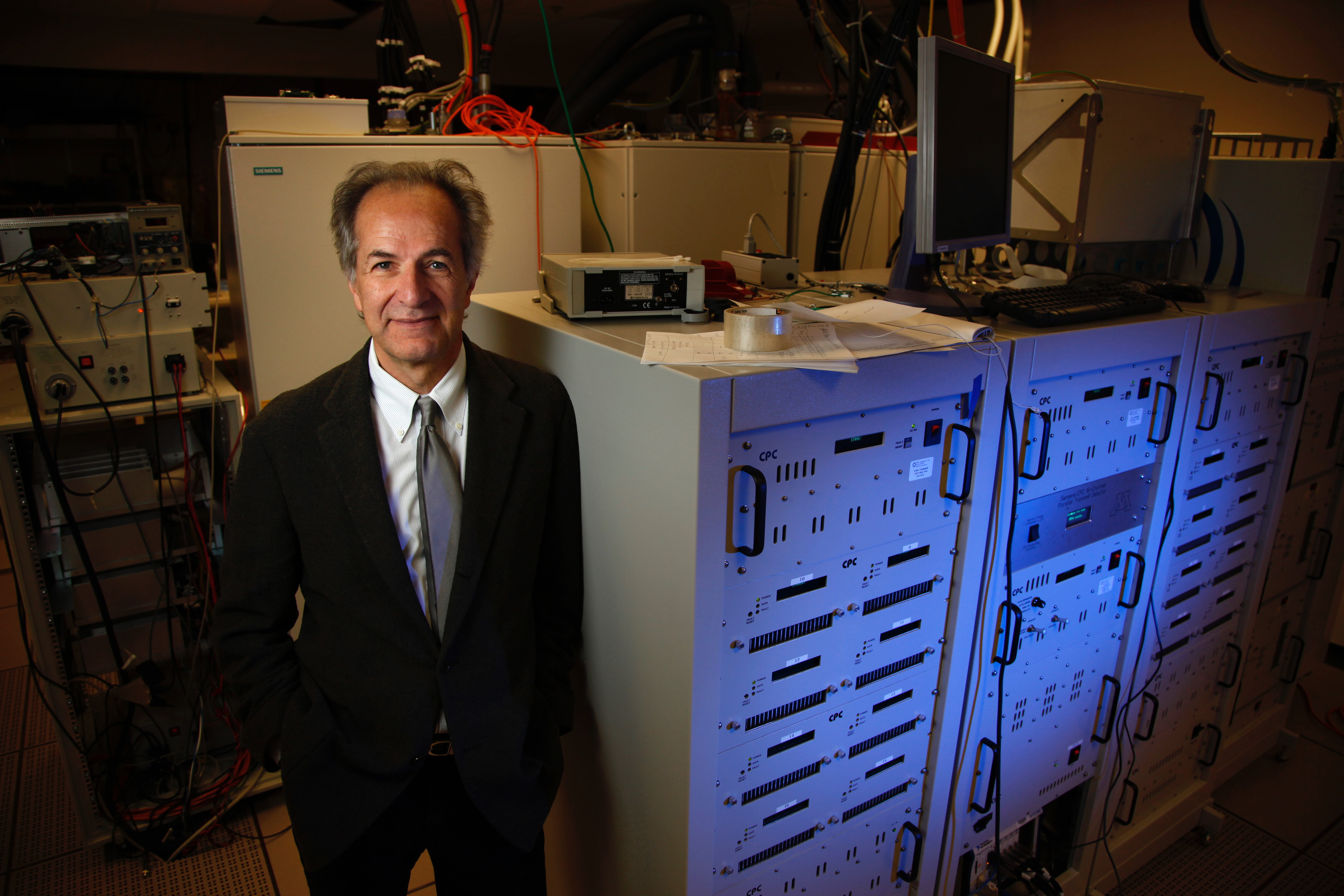
Kamil Ugurbil, Ph.D.
|
|
||||||||
Kamil Ugurbil currently holds the McKnight Presidential Endowed Chair Professorship in Radiology, Neurosciences, and Medicine and is the Director of the Center for Magnetic Resonance Research (CMRR) at the University of Minnesota. Prof. Ugurbil was educated at Robert Academy, Istanbul (high school) and Columbia University, New York, N.Y. After completing his B.A. and Ph.D. degrees in physics, and chemical physics, respectively, at Columbia, he joined AT&T Bell Laboratories in 1977, and subsequently returned to Columbia as a faculty member in 1979. He was recruited to the University of Minnesota in 1982 where his research in magnetic resonance led to the evolution of his laboratory into an interdepartmental and interdisciplinary research center, the CMRR.
The work that introduced magnetic resonance imaging of neuronal activity in the human brain (known as fMRI) was accomplished independently and simultaneously in two laboratories, one of which was Ugurbil's in CMRR. Since then, his primary focus has been the development and application of methods capable of obtaining high resolution and high accuracy functional and anatomical information in the human brain, targeting spatial scales ranging from the whole brain to elementary neuronal ensembles exemplified by cortical columns and layers. This body of work has culminated in unique accomplishments, such as the first time functional mapping of orientation columns in the human primary visual cortex, and involved the development of extensive new instrumentation and image acquisition approaches, including the introduction and development of ultrahigh magnetic fields (7 Tesla and higher) for functional and anatomical imaging and highly accelerated functional brain imaging. This body of work was recognized by several awards and honors including:
- IEEE Medal for Innovations in Healthcare Technology
- Koç Award
- Richard Ernst Lecture and Gold Medal
- Election to the National Academy of Medicine
- Election to the American Academy of Arts and Sciences (USA)
- Election to the National Academy of Inventors (USA)
- Membership in NIH Working Group on Brain Research through Advancing Innovative Neurotechnologies (BRAIN) Initiative
- Honorary Doctorate (Doctorate Honoris Causa), University of Maastricht, Netherlands
- Honorary Doctorate (Doctorate Honoris Causa), University of Utrecht, Netherlands
- Gold Medal, International Society of Magnetic Resonance in Medicine (ISMRM)
- Co-Principal Investigator, Human Connectome Project (http://humanconnectome.org/)
- Membership in National Institute of Mental Health (NIMH), Board of Scientific Advisors
- Irma T. Hirschl Career Scientist Award
- Hammett Award for Original and Distinguished Research
- Columbia University, Graduate Faculties Alumni Scholar
Research Focus
- Imaging brain function and connectivity with magnetic resonance (MR) techniques at ultrahigh magnetic fields (7 Tesla and higher)
- Mechanisms underlying functional mapping signals in fMRI
- Development of biological MR imaging and spectroscopy methods at ultrahigh magnetic fields for biomedical research in the human body (the first 7 Tesla instrument for human studies was developed in CMRR in 1999)
- Development of high frequency RF instrumentation for ultrahigh field MR imaging in humans
Video of Prof. Ugurbil receiving the Gold Medal and delivering the Ernst Lecture
https://www.chab.ethz.ch/en/research/institutes/LPC/ernst-lecture/2014.html
20140520_HGF30_Richard_Ernst_Lecture_Ugurbil.mp4
Selected Publications and Contributions to Science
1. Discovery of Functional Magnetic Resonance Imaging (fMRI). The introduction of fMRI was accomplished in two laboratories independently and simultaneously in 1991-1992, one of which was Ugurbil's at the University of Minnesota. Using manipulations of the physiologic state of the anesthetized animal, such as altering oxygen content of inhaled gas, S. Ogawa from Bell Laboratories described in 1990 the effect of deoxyhemoglobin on MR images of the brain, and named it Blood Oxygenation Dependent (BOLD) contrast. The use of this contrast mechanism for functional mapping of human brain activity was achieved in Ugurbil’s laboratory in collaboration with Ogawa. This landmark effort was also accompanied by the first modeling and experimental papers aimed at elucidating the mechanism underlying the functional imaging signals.
- Ogawa S, Tank DW, Menon R, Ellermann JM, Kim SG, Merkle H, Ugurbil K. (1992). “Intrinsic signal changes accompanying sensory stimulation: functional brain mapping with magnetic resonance imaging.” Proc Natl Acad Sci U S A 89(13): 5951-5955. PMCID: PMC402116.
- Ogawa S, Menon RS, Tank DW, Kim SG, Merkle H, Ellermann JM, Ugurbil K. (1993). “Functional brain mapping by blood oxygenation level-dependent contrast magnetic resonance imaging. A comparison of signal characteristics with a biophysical model.” Biophys J 64(3): 803-812. PMCID: PMC1262394.
- Menon RS, Ogawa S, Tank DW, Ugurbil K. (1993). “4 Tesla gradient recalled echo characteristics of photic stimulation-induced signal changes in the human primary visual cortex.” Magn Reson Med 30(3): 380-386.
- Kim SG, Hendrich K, Hu X, Merkle H, Ugurbil K. (1994). "Potential pitfalls of functional MRI using conventional gradient-recalled echo techniques". NMR Biomed;7(1-2):69-74
- Kim S-G, Richter W, Ugurbil K. (1997). "Limitations of temporal resolution in functional MRI". Magn Reson Med; 37(4):631-636.
2. Understanding mechanisms underlying functional mapping signals in fMRI; towards developing high resolution and high accuracy maps of neuronal activity. The ability to obtain functional maps at the level of minimal architectural units that organize neural populations of similar properties is critical for understanding brain function. The cortical columns of neocortex are prominent examples of such structurally and functionally specialized units and have received extensive attention in studies of brain function using electrophysiology, optical imaging, and computational modeling. In addition, the differences in connectivity and cell types across the few millimeter thick cortical ribbon imply that laminar resolution is also critical in deciphering the elementary computations of the brain. However, because fMRI signals reflect neuronal activity indirectly through neurovascular coupling and vasculature, it is not possible to assume a priori that functional mapping signals in fMRI have high fidelity to sites of neuronal activity. Subsequent to the introduction of fMRI, Ugurbil’s group made seminal and pioneering contributions towards understanding the mechanisms underlying fMRI signals, the spatial scale of neurovascular coupling, and the nature of mapping signal with different functional contrast encoding approaches; this knowledge was then exploited to develop methods (including ultrahigh field MR technology (see below)) for functional mapping at the level of cortical columns and layers in the human brain.
- Duong TQ, Kim DS, Ugurbil K, Kim SG. (2001). "Localized cerebral blood flow response at submillimeter columnar resolution." Proc Natl Acad Sci U S A 98, 10904-10909. PMCID: PMC58572.
- Shmuel A, Yacoub E, Pfeuffer J, Van de Moortele PF, Adriany G, Hu X, Ugurbil K. (2002). "Sustained negative BOLD, blood flow and oxygen consumption response and its coupling to the positive response in the human brain." Neuron 36, 1195-1210.
- Shmuel A, Yacoub E, Chaimow D, Logothetis NK, Ugurbil K. (2007). "Spatio-temporal point-spread function of fMRI signal in human gray matter at 7 Tesla." Neuroimage 35, 539-552. PMCID: PMC2989431.
- Yacoub E, Harel N, Ugurbil K. (2008). "High-field fMRI unveils orientation columns in humans." Proc Natl Acad Sci U S A 105, 10607-10612. PMCID: PMC2492463.
- Uludag K, Muller-Bierl B, and Ugurbil K. (2009). “An Integrative Model for Neuronal Activity-Induced Signal Changes for Gradient and Spin Echo Functional Imaging.” Neuroimage, 2009. 48(1): p. 150-65.
- De Martino F, Moerel M, Ugurbil K, Goebel R, Yacoub E, and Formisano E. (2015). “Frequency Preference and Attention Effects across Cortical Depths in the Human Primary Auditory Cortex”. Proc Natl Acad Sci U S A, 2015. DOI: 10.1073/pnas.1507552112.
3. Development of high and ultrahigh magnetic fields for magnetic resonance imaging and spectroscopy. Motivated primarily by the understanding of the mechanisms underlying functional mapping signals in fMRI and the role played by the static magnetic field strength, a common thread in Ugurbil’s work has been the effort to exploit very high magnetic fields for human studies in order to enhance the biological information content, accuracy, and resolution of imaging and spectroscopy signals. Ugurbil laboratory was one of the first three academic laboratories that initiated 4 Tesla (T) human imaging at approximately the same time in ~1990; subsequently, justified by a large body of 4T human data and small animal experiments conducted at 9.4T, Ugurbil and colleagues were the first to introduce 7 Tesla for studies of human brain function in ~1999, using a 7T magnet developed specifically the first time for this effort and with system development and integration undertaken by Ugurbil and colleagues. This seminal effort in ultrahigh magnetic fields was complemented with fundamental studies on the physics of high field/high frequency imaging in the human body, development of high frequency RF methods and instrumentation (such as parallel transmit concepts and hardware), and introduction of new data acquisition methods, to attain some of the most advanced neuroimaging capabilities. The data coming from this 7T system ultimately led to commercially produced 7T systems and to the evolution of such high fields as the most advanced neuroimaging and more recently body imaging platform.
- Ugurbil K, Garwood M, Ellermann J, Hendrich K, Hinke R, Hu X, Kim SG, Menon R, Merkle H, Ogawa S, Salmi R. (1993). “Imaging at high magnetic fields: initial experiences at 4 T”. Magn Reson Q 9, 259-277.
- Vaughan JT, Garwood M, Collins CM, Liu W, DelaBarre L, Adriany G, Andersen P, Merkle H, Goebel R, Smith MB, Ugurbil K. (2001). “7T vs. 4T: RF power, homogeneity, and signal-to-noise comparison in head images.” Magn Reson Med 46, 24-30.
- Adriany G, Van de Moortele PF, Wiesinger F, Moeller S, Strupp JP, Andersen P, Snyder C, Zhang X, Chen W, Pruessmann KP, Boesiger P, Vaughan T, Ugurbil K. (2005). “Transmit and receive transmission line arrays for 7 Tesla parallel imaging.” Magn Reson Med 53, 434-445.
- Van de Moortele PF, Akgun C, Adriany G, Moeller S, Ritter J, Collins CM, Smith MB, Vaughan JT, Ugurbil K. (2005). “B(1) destructive interferences and spatial phase patterns at 7T with a head transceiver array coil.” Magn Reson Med 54, 1503-1518.
- Ugurbil K. (2012). "The road to functional imaging and ultrahigh fields". Neuroimage;62(2):726-735.
- Ugurbil K. (2014). “Magnetic resonance imaging at ultrahigh fields”. IEEE Trans Biomed Eng;61(5):1364-1379.
4. The Human Connectome Project. The Human Connectome Project (HCP) was a major undertaking funded by the sixteen institutes and centers of the National Institutes of Health (NIH) that support the NIH Blueprint for Neuroscience Research. HCP aims to map connections in of the human brain in the mm scale in normal adults in their mid-life. This project was awarded to a consortium led by the Washington University and the University of Minnesota, Center for Magnetic Resonance Research (CMRR) (grant number 1U54MH091657) with David Van Essen from Washington University in St. Louis and Ugurbil serving as co-PIs. Ugurbi’s group was responsible for all the technical developments for image acquisition and reconstructions methods. Starting from developments already in progress in Ugurbil group, major advances for image acquisition were accopmlished leading to the highest temporal and spatial resolution fMRI and diffusion weighted (dMRI) images of the human brain. These imaging approaches have redefined functional and diffusion weighted imaging. Tom Insel, the head of the NIMH, cited the HCP accomplishments as a major advance in brain sciences (http://www.nimh.nih.gov/about/director/2015/brain-awareness.shtml).
- Ugurbil K, Xu J, Auerbach EJ, Moeller S, Vu AT, Duarte-Carvajalino, et al. (2013). “Pushing spatial and temporal resolution for functional and diffusion MRI in the Human Connectome Project.” Neuroimage 80, 80-104.
- Van Essen DC, Smith SM, Barch DM, Behrens TE, Yacoub E, Ugurbil K, WU-Minn HCP Consortium. (2013). “The WU-Minn Human Connectome Project: An overview.” Neuroimage 80, 62-79.
- Smith SM, Miller KL, Moeller S, Xu J, Auerbach EJ, Woolrich MW, Beckmann CF, Jenkinson M, Andersson J, Glasser MF, Van Essen DC, Feinberg DA, Yacoub ES, Ugurbil K. (2012). “Temporally-independent functional modes of spontaneous brain activity.” Proc Natl Acad Sci U S A 109, 3131-3136.
- Xu J, Moeller S, Auerbach EJ, Strupp J, Smith SM, Feinberg DA, Yacoub E, Ugurbil K. 2013. "Evaluation of slice accelerations using multiband echo planar imaging at 3 T." Neuroimage 83, 991-1001.
- Vu AT, Auerbach E, Lenglet C, Moeller S, Sotiropoulos SN, Jbabdi S, Andersson J, Yacoub E, Ugurbil K. “High resolution whole brain diffusion imaging at 7T for the Human Connectome Project”. Neuroimage 2015;122:318-331.
5. In vivo magnetic resonance spectroscopy and applications to studies in the human brain. One of the first pioneering efforts towards using magnetic resonance to extract biochemical and physiologic information non-invasively in intact biological systems was started in Bell Laboratories Biophysics group where Ugurbil worked after his PhD. In this small group, Ugurbil and colleagues introduced and demonstrated the use of magnetic resonance spectroscopy in intact biological systems. Many years later many of these techniques were used to study bioenergetics of neuronal function in the human brain at ultrahigh magnetic fields in Ugurbil’s laboraty in the Iniversity of Minnesota.
- Ugurbil K, Brown TR, den Hollander JA, Glynn P, Shulman RG. (1978). "High-resolution 13C nuclear magnetic resonance studies of glucose metabolism in Escherichia coli." Proc Natl Acad Sci U S A 75, 3742-3746.
- Brown TR, Kincaid BM, Ugurbil K. (1982). "NMR chemical shift imaging in three dimensions." Proc Natl Acad Sci U S A 79, 3523-3526.
- Ugurbil K, Rottenberg H, Glynn P, and Shulman R G. (1982). "Phosphorus-31 nuclear magnetic resonance studies of bioenergetics in wild-type and adenosinetriphosphatase(1-) Escherichia coli cells." Biochemistry 21(5): 1068-1075.
- Gruetter R, Weisdorf SA, Rajanayagan V, Terpstra M, Merkle H, Truwit CL, Garwood M, Nyberg SL, Ugurbil K. (1998). "Resolution improvements in in vivo 1H NMR spectra with increased magnetic field strength". J Magn Reson;135(1):260-264.
- Chen W, Zhu XH, Gruetter R, Seaquist ER, Adriany G, Ugurbil K. (2001). "Study of tricarboxylic acid cycle flux changes in human visual cortex during hemifield visual stimulation using (1)H-{(13)C} MRS and fMRI." Magn Reson Med 45, 349-355.
- Mangia S, Tkac I, Gruetter R, Van de Moortele PF, Maraviglia B, Ugurbil K. (2007). "Sustained neuronal activation raises oxidative metabolism to a new steady-state level: evidence from 1H NMR spectroscopy in the human visual cortex." J Cereb Blood Flow Metab 27, 1055-1063.
6. Design of novel RF pulses, and MR acquisition sequences. Development of new methods for MR image or spectra acquisitions and radio-frequency (RF) pulse design, and improvements of such methods has been an integral part of Ugurbil’s work from the very beginning of his career. Some of the most commonly used methods in the biomedical applications of MR were introduced by Ugurbil and his colleagues and include, for example, chemical shift imaging for spectroscopy; adiabatic “plane-rotation” RF pulses, their optimization, and their use in spatial-spectroscopic encoding; parallel transmit methods for RF pulse design for improved RF homogenity and power deposition constraint; high contrast anatomical imaging at high magnetic fields; and most notably for recent efforts to study the human brain, the introduction of slice accelerated multiband (MB), simultaneous multislice (SMS) imaging for fMRI and diffusion imaging (for tractography), which has transformed the way studies of human brain connectivity and function is currently performed. Few examples of publications that cover these efforts include:
- Brown TR, Kincaid BM, Ugurbil K. (1982). "NMR chemical shift imaging in three dimensions". Proc Natl Acad Sci U S A;79(11):3523-3526.
- Ugurbil K, Garwood M, Rath A, Bendall MR. (1988). "Amplitude and Frequency/Phase Modulated Refocusing Pulses that Induce Plane Rotations Even in the Presence of Inhomogeneous Fields". J Magn Reson;78:472-497.
- Merkle H, Wei H, Garwood M, Ugurbil K. (1992). "B1-Insenstive Heteronuclear Adiabatic Polarization Transfer for Signal Enhancement". J of Magn Reson;99:480-494.
- Lee JH, Garwood M, Menon R, Adriany G, Andersen P, Truwit CL, Ugurbil K. (1995). "High contrast and fast three-dimensional magnetic resonance imaging at high fields". Magn Reson Med;34(3):308-312.
- Moeller S, Yacoub E, Olman CA, Auerbach E, Strupp J, Harel N, Ugurbil K. (2010). "Multiband multislice GE-EPI at 7 tesla, with 16-fold acceleration using partial parallel imaging with application to high spatial and temporal whole-brain fMRI". Magn Reson Med;63(5):1144-1153.
- Feinberg DA, Moeller S, Smith SM, Auerbach E, Ramanna S, Gunther M, Glasser MF, Miller KL, Ugurbil K, Yacoub E. (2010). "Multiplexed echo planar imaging for sub-second whole brain FMRI and fast diffusion imaging". PLoS ONE;5(12):e15710.
- Auerbach EJ, Xu J, Yacoub E, Moeller S, Ugurbil K. (2013). "Multiband accelerated spin-echo echo planar imaging with reduced peak RF power using time-shifted RF pulses". Magn Reson Med;69(5):1261-1267.
- Wu X, Schmitter S, Auerbach EJ, Moeller S, Ugurbil K, Van de Moortele PF. (2013). "Simultaneous multislice multiband parallel radiofrequency excitation with independent slice-specific transmit B1 homogenization". Magn Reson Med;70(3):630-638.
