
CMRR
Center for Magnetic Resonance Research, Department of Radiology
News
You are here
A powerful force
Jan 01, 2009 The Center for Magnetic Resonance Research, soon to house the world’s strongest magnet, is pushing the technology’s limits.
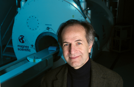
Some scientists make strides in biomedical research by acquiring state-of-the-art equipment and then using it to answer questions about living systems. “Good research can be done in that fashion,” says Kamil Ugurbil, Ph.D., director of the University of Minnesota’s Center for Magnetic Resonance Research (CMRR).
But Ugurbil takes a different approach. A chemical physicist by training, he has always veered away from using new machines straight out of the box. Instead, he likes to develop novel technologies and pushes them beyond what anyone ever imagined they could do. Even at points when other experts in the field believed magnetic resonance tools had reached their limits, Ugurbil and his colleagues have persisted, extending the capabilities of the magnets and finding new applications for them.
He puts it this way: “We’re excited when we can get information that is beyond the bread and butter of the technology.” That desire to test the untapped potential of new high-field magnets has placed the CMRR among world leaders in imaging. It’s also stretched every parameter of the discipline.
Today the burgeoning center, which Ugurbil has led since 1991, has 21 faculty members and six high-field magnets, with an additional “ultra-high-field” magnet on the way. As the latest construction project in the University’s developing research park, called the Biomedical Discovery District, the CMRR building is undergoing a renovation and large-scale expansion expected to be completed by fall 2010. A $53 million budget, provided in part by the state, will add approximately 65,000 square feet for new research and clinical studies.
The expanded building will house a new magnet that will be the highest field ever attained for human studies. (The magnets are referred to by the strength of their magnetic fields; typical magnetic resonance imaging [MRI] machines used for hospital diagnoses have a 1.5 Tesla magnet.) And an astounding 16.4 Tesla magnet currently being installed in the renovated section of the building will be the largest magnet in the country; the only other one of its kind is in Europe. These technologies are so new that even Ugurbil isn’t certain what they’ll be capable of revealing, although his team’s track record with new tools suggests that remarkable discoveries are on the horizon.
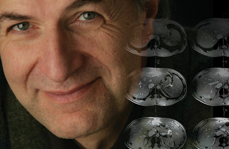
Center for Magnetic Resonance Research director Kamil Ugurbil, Ph.D., develops novel imaging technologies and pushes the boundaries of what they can do. The 9.4T magnet pictured behind Dr. Kamil Ugurbil is part of the world's highest-field magnetic resona
Center for Magnetic Resonance Research director Kamil Ugurbil, Ph.D., develops novel imaging technologies and pushes the boundaries of what they can do. The 9.4T magnet pictured behind Dr. Kamil Ugurbil is part of the world's highest-field magnetic resona
A paradigm shift
The changing geographic location of magnetic resonance (MR) research on the University campus tells the story about the rising promise and prominence of Ugurbil’s group. When Ugurbil arrived at the University of Minnesota in 1978 after working at Bell Laboratories and then at Columbia University, his lab was housed with the Gray Freshwater Biological Institute, located on the St. Paul campus, far from the University’s medical center. (The institute is no longer part of the University.)
At Bell Labs, Ugurbil had written papers about applying MR spectroscopy to cellular metabolism that would shortly become classics. At the University, he intended to continue exploring what magnetic resonance technology could do with cells and even intact organs.
But then, with a program project grant from the National Institutes of Health and matching funds from the University’s administration, the Freshwater Biological Institute obtained a 4.7 Tesla magnet with a bore capable of holding small animals. Ugurbil’s group began to think about studying intact living organisms with the high-field magnet.
“We had the basis to believe we could succeed in that area,” Ugurbil says. The magnet was so new that the group had to develop its own software applications to use it. But as they succeeded in attaining useful images of rat organs, they became intrigued by a bigger challenge: applying the technology to humans.
It turns out that humans are complicated subjects for MR studies. Both building the high-field magnet to accommodate a person and creating an image as the magnetic fields increase become more difficult. In the late 1980s, no groups anywhere were having success with increased field strength for human imaging.
“It was accepted at the time that 1 to 1.5 [Tesla] would be the optimal magnetic field for doing MR research and for clinical diagnoses,” says Michael Garwood, Ph.D., associate director of the CMRR, who was then a postdoc at the University.
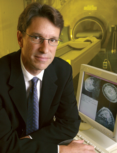
Brain scans performed by cancer researcher Michael Garwood, Ph.D., in the late 1980s showed the advantage of high-resolution 4 Tesla images over 1 or 1.5 Tesla, then thought to be the optimal magnetic field for research and clinical use.
Higher and higher
But keen on improving the magnets’ resolution and sensitivity, Ugurbil linked his group with the Medical School’s radiology department. Bill Thompson, M.D., the department chair at the time and an ardent supporter of advancing MR research—aided by David Brown, M.D., who was dean of the Medical School—enabled Ugurbil to acquire one of the first industry-built 4 Tesla systems. The instrument was set up in the CMRR’s first home, a building near the University’s medical center on East River Road.
Acquiring that magnet involved a leap of faith, Ugurbil acknowledges today. Even industry members had abandoned the idea of developing a 4 Tesla magnet to study humans; the images it produced were less clear than a standard magnetic resonance image. But Ugurbil believed the magnet’s higher sensitivity offered an opportunity to map brain activity, and his group developed strategies to address some of the confounding factors.
He and his team forged ahead with plans to use the magnet to study increased oxygenation in areas of the brain. In collaboration with Bell Laboratories, they mapped active neuronal regions of the brain in living subjects, a technique known as functional imaging or fMRI. At the same time, Garwood demonstrated that, contrary to expectations at the time, it was feasible to obtain beautiful anatomical images of the human brain at 4 Tesla.
“The very first experiments we did on the 4 Tesla were great successes,” recalls Ugurbil. They offered a paradigm shift in how MR could be used.
At the time, along with the 4 Tesla machine, the group had two magnets for in vivo animal studies and continued to advance MR spectroscopy. But with little room to expand, and with an interest in attaining new magnets, in 1998 the CMRR moved into its current building, a low, brightly lit structure that helps anchor the emerging research park on the north side of the University’s new TCF Bank Stadium.
Over the next decade, the group acquired an array of new magnets for both animal and human research, and the building underwent three separate renovations to accommodate them. The 7 Tesla, 90 cm bore magnet the group brought online in 1999 was the world’s first of its kind developed for human studies. This was made possible by major support from the W. M. Keck Foundation, the University of Minnesota, the National Science Foundation, and the National Institutes of Health.
In 2002, a $4.5 million gift from the W. M. Keck Foundation was instrumental in developing a unique 9.4 Tesla, 65 cm bore imaging system capable of accommodating human studies, again the world’s first. Each of these steps to higher magnetic fields has yielded major gains in sensitivity, image resolution and specificity (for functional imaging), and chemical resolution—the ability to distinguish neurochemicals from one another (using MR spectroscopy).
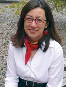
Biochemist Gülin Öz, Ph.D., is using magnetic resonance spectroscopy to track chemical precursors to spinocerebellar ataxia with the hopes of disrupting the process before the condition causes irreversible damage to the brain.
Focused on the brain
For University researchers from a variety of disciplines, the collection of magnets and the in-house expertise at the Center for Magnetic Resonance Research offer unparalleled opportunities to study diseases. With funding from the National Institute of Mental Health, University psychiatry professor Kelvin Lim, M.D., for example, has collected magnetic resonance images of anatomical differences in the brains of people with schizophrenia. In particular, he’s looking at the gray matter deficit that occurs early during the disease course.
At the same time, he’s been able to measure changes in brain activity using fMRI. The studies so far have looked at humans in the CMRR’s 3 Tesla magnet, but Lim hopes to develop techniques to use the center’s even higher-resolution 9.4 Tesla magnet to do magnetic resonance spectroscopy, which will offer a glimpse of specific neurochemical changes in schizophrenic patients.
“Access to this type of hardware gives us a tremendous advantage here,” he says. Ultimately, these refined views of the brain may reveal subcategories of schizophrenia, each with its own unique characteristics. “One of my goals is to get to the point,” Lim says, “where we have important biomarkers [for each subcategory] that can help us guide treatment.”
Down the hall, CMRR biochemist Gülin Öz, Ph.D., is studying the effects on the brain of a mutant gene that causes a neurodegenerative disease known as spinocerebellar ataxia. In humans, where her MR investigations began, the genetic condition eventually results in irreversible damage to the cerebellum, causing movement problems like loss of balance and coordination as well as awkward gait. But as she sought more specific and controlled biochemical information about what was happening to the cells, Öz redirected her MR spectroscopy studies to look at the brains of mice that have the mutation.
In collaboration with ataxia expert Harry Orr, Ph.D., in the Department of Laboratory Medicine and Pathology and Institute of Human Genetics, who provided a mouse model for the disease, Öz is conducting research to pinpoint chemical precursors to the devastating structural changes that the neurons undergo.
“It’s been shown that if you can intervene early on in the disease, you can reverse the changes and rescue the cells before they die,” Öz explains. What’s more, better understanding of the neurochemistry may reveal commonalities among a range of neurodegenerative disorders, providing information about other diseases like Alzheimer’s and Parkinson’s.
Other projects delve into different aspects of various disease processes, from a study taking place with the Mayo Clinic that looks at the amyloid plaques that form in people with Alzheimer’s disease (researchers at the CMRR were the first ever to visualize those plaques in living organisms) to a collaboration with University of Minnesota diabetes researcher Elizabeth “Betsy” Seaquist, M.D., who is investigating how diabetic “sugar crashes” affect metabolism in the brain.
A $7.9 million grant from the National Institutes of Health in 2006 helps ensure that the CMRR’s state-of-the-art equipment is available to these and other neurosciences researchers across the University.
An unprecedented resource
Now, with sights set on bringing in new ultra-high-field magnets, the CMRR faces new research possibilities—and new challenges. Even though the 16.4 Tesla magnet for studying animals is powerful enough to visualize a process as minute as the development of a rat embryo, it may take years to develop the methodologies that can take full advantage of the magnet’s capabilities. In typical style, Ugurbil pushed to acquire the tools to get that work started. (The project received University support as well as $2 million from the NIH.)
What’s clear is that the work Ugurbil began 25 years ago has created an unprecedented resource. “There’s no setup quite like the one we have here, in scope of instrumentation and quality of leadership,” states Charles Moldow, M.D., vice dean for research and operations in the Medical School.
The construction planned for the research park around the CMRR will situate the building among other medical research as it’s never been positioned before, notes Kevin Ross, capital planning project manager for the expansion. New skyways linking the building to its neighbors will connect it with labs in such fields as neuroscience, immunology, and stem cell research. Until now, the building has “stood very much isolated from campus, kind of on the outskirts,” says Ross. “Now, when the CMRR expansion is completed, it will be in the heart of a state-of-the-art research community.”
By Kate Ledger