
CMRR
Center for Magnetic Resonance Research, Department of Radiology
Research Highlights
You are here
Brain Angiography
|
|
|
3D Time of flight (TOF) imaging is the most clinically used non-contrast enhanced magnetic resonance angiography (MRA) technique to visualize vessels and blood flow in the human brain, in particular for the arteries. Compared to other common techniques in clinical route TOF is a non-invasive method and patients are not exposed to ionizing radiation. The method is increasingly used in clinics for acute stroke imaging and also to detect cerebral aneurisms, stenosis and other cerebral diseases.
TOF contrast and image quality improves significantly with increasing magnetic field strength of the MRI scanner. Thus at CMRR we perform TOF imaging at ultra high field (UHF) with more than twice the clinical field strength to investigate its potential for future clinical applications. However, with increasing field strength more energy is required to acquire the MR images, which inhibits to transfer commonly used TOF imaging strategies from clinical field strength to UHF. Thus important modules used standardly in clinical TOF to improve the contrast or to selectively visualize the arteries (without the veins) have to be skipped in UHF TOF.
At CMRR we have modified the image acquisition process to reduce the energy needed for UHF angiogram images. By this we were able to acquire TOF angiograms including all clinical modules and we could successfully demonstrate a contrast improvement of more than 50% by using the additional modules. Figure 1b shows the improvement on a transversal angiogram image acquired at UHF compare to the Figure 1a where the additional modules were skipped. With this technique we are able to generate high resolution angiograms with significantly higher contrast and image quality compared to clinical acquisitions.
We believe that the results are encouraging to bring ultra high field TOF closer towards clinical application, which may help to more accurately detect brain pathologies.
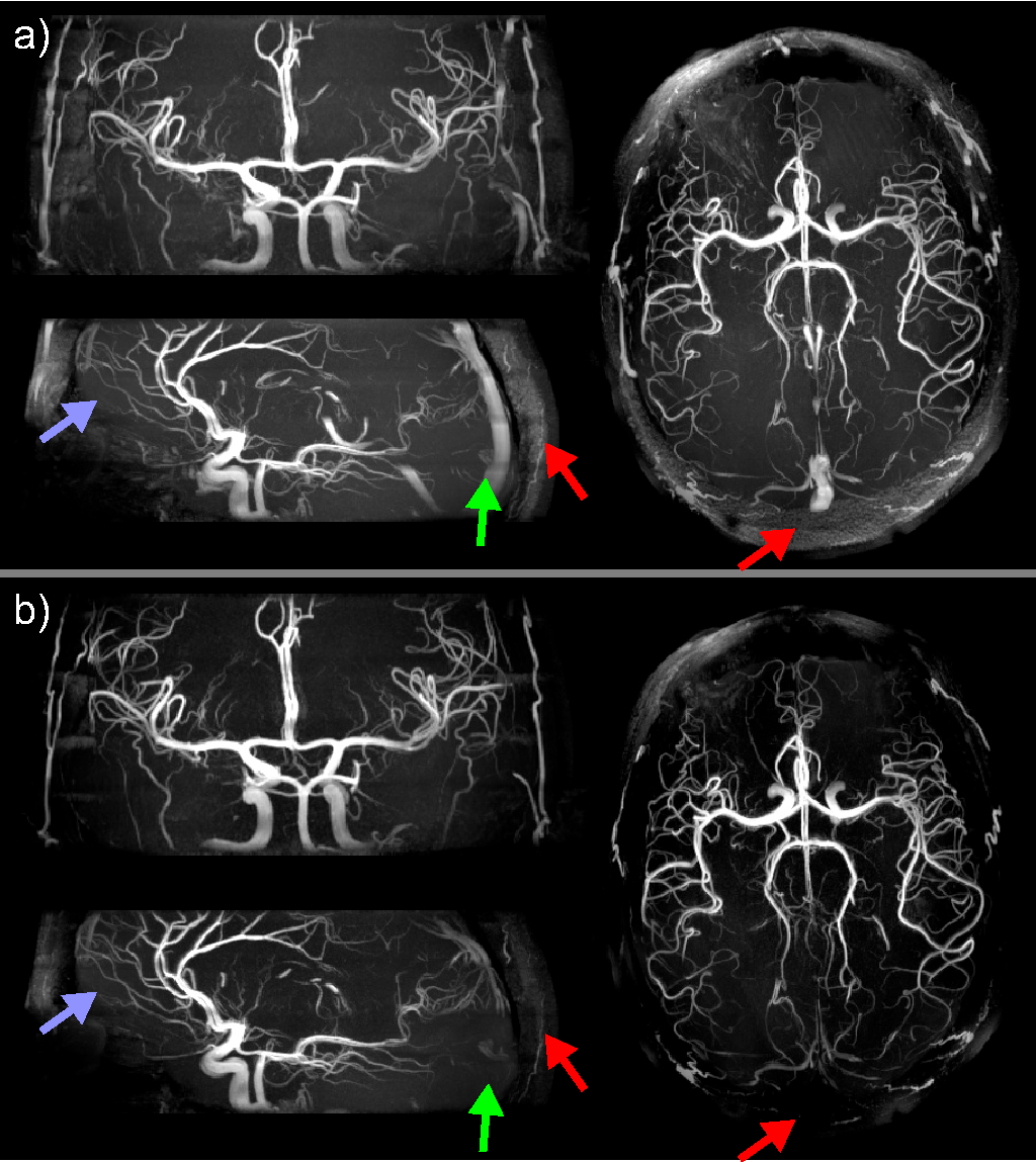
Angiographic views from different orientations of the standard acquisition without (a) and with (b) additional TOF modules, acquired at ultra high field. Due to the improve contrast in b smaller vessels become visible more clearly, especially in the frontal areas of the brain (blue arrow). To visualize only the cerebral arteries, venous signal is efficiently suppressed in b compared to a (green arrow). In addition the subcutaneous fat signal is strongly reduced (red arrows) in b.