
CMRR
Center for Magnetic Resonance Research, Department of Radiology
OPTIMAL FAIR Sequence
You are here
OPTIMAL FAIR
Background
Arterial spin labeling (ASL) measurements of blood flow in human hippocampus are complicated by its relatively small size and unusual shape. The diameter of the human hippocampus tapers posteriorly along its 45–50-mm curved length from a relatively wide head (8–12 mm) to a thinner body (6–8 mm) and even thinner tail (2–6 mm). The coarse spatial resolution (isotropic 3.5-mm voxels) of standard ASL imaging limited the number of voxels that could be sampled without partial volume effects where the narrow aspects of the body and tail of the hippocampus curved through the slice planes, hindering the ability to conclusively characterize and distinguish middle and posterior regions. To overcome these limitations, a standard FAIR (Flow-sensitive Alternating Inversion Recovery) pulse sequence was modified by rotating the imaging slice plane orthogonal to the oblique axial plane of the tagging inversion. With this Orthogonally Positioned Figure 1 Spatial definitions of different slabs Tagging Imaging Method for Arterial Labeling with FAIR (OPTIMAL FAIR) technique, oblique coronal imaging slices with high in-plane resolution can be placed perpendicular to the longitudinal axis of the hippocampus along the A–P direction (Figure 1). This reduces partial volume effects and increases resolution, allowing the detection of finer details of the A–P differences in perfusion parameters, thus enabling reliable studies of these perfusion differences in both normal and compromised or diseased hippocampus (1-3).
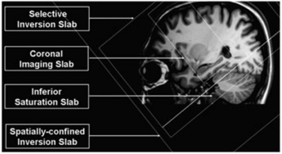
Figure 1. Spatial definitions of different slabs in Orthogonally Positioned Tagging Imaging Method for Arterial Labeling with Flow-sensitive Alternating Inversion Recovery (OPTIMAL FAIR) (1).
Features
- The OPTIMAL FAIR sequence contains a sequence module for temporal bolus width definition by using either QUIPSS II or Q2TIPS methods, and these two methods can also be turned off when the number of saturation pulses (via user interface (UI) parameter “QSAT.No.”) is set to zero.
- Imaging slab pre-saturation is supported with two execution modes: a single saturation before ASL inversion RF pulses or one pre-inversion saturation combined with two post-inversion saturation and minimize the subtraction errors caused by the imperfect inversion profile (4).
- HSN RF pulse (N>= 4) can be selected for FAIR inversions to reduce RF peak power that is usually limited at ultra high fields, e.g. 7T.
- Both QUIPSS II and Q2TIPS are supported with great flexibility via multiple UI parameters: the number, duration, slab size and temporal gaps of saturation RF (please refer Figure 1 in the reference 4).
- Up to two M0 images can be acquired at the end of ASL series acquisition.
- Bi-polar flow-encoding/crushing gradients are available to suppress hyperintense intravascular signals.
CMRR C2P
If you are interested in obtaining these sequences, the CMRR C2P instructions and agreement can be found on this page at the UMN Office for Technology Commercialization. Once you have executed a C2P agreement and have been given an access password, the sequence binaries can be downloaded here.
Note to users: If you have noticed a bug or have a request for a new feature in a future release, please email Xiufeng Li lixx1607@umn.edu to open a new support request. Be sure to include the sequence variant and the model of scanner you are using in the problem description.
Download OPTIMAL FAIR Sequence:
|
OPTIMAL FAIR Release 1.0 |
|
|
Released date OPTIMAL FAIR Release Notes v1.0 |
Download coming soon
 Release 1.0 for VB17 (build ) Release 1.0 for VB17 (build ) |
References
1. Li X, Sarkar SN, Purdy DE, Spence JS, Haley RW, Briggs RW. Anterioposterior perfusion heterogeneity in human hippocampus measured by arterial spin labeling MRI. NMR Biomed. 2013 Jun; 26(6):613-21. doi: 10.1002/nbm.2898.
2. Li X, Spence JS, Buhner DM, Haley RW, Briggs RW. Dynamic physostigmine effects on hippocampus perfusion. J Magn Reson Imaging. 2012;35(2):280-286; doi: 10.1002/jmri.22821.
3. Li X, Spence JS, Buhner DM, Hart J, Jr., Cullum CM, Biggs MM, Hester AL, Odegard TN, Carmack PS, Briggs RW, Haley RW. Hippocampal dysfunction in Gulf War veterans: investigation with ASL perfusion MR imaging and physostigmine challenge. Radiology 2011;261(1):218-225.
4. Li X, Sarkar SN, Purdy DE, Haley RW, Briggs RW. Improved quantification of brain perfusion using FAIR with active suppression of superior tagging (FAIR ASST). J Magn Reson Imaging. 2011;34(5):1037-1044; doi: 10.1002/ jmri.22734.