
CMRR
Center for Magnetic Resonance Research, Department of Radiology
Multi-band EPI PCASL Sequence
You are here
Background
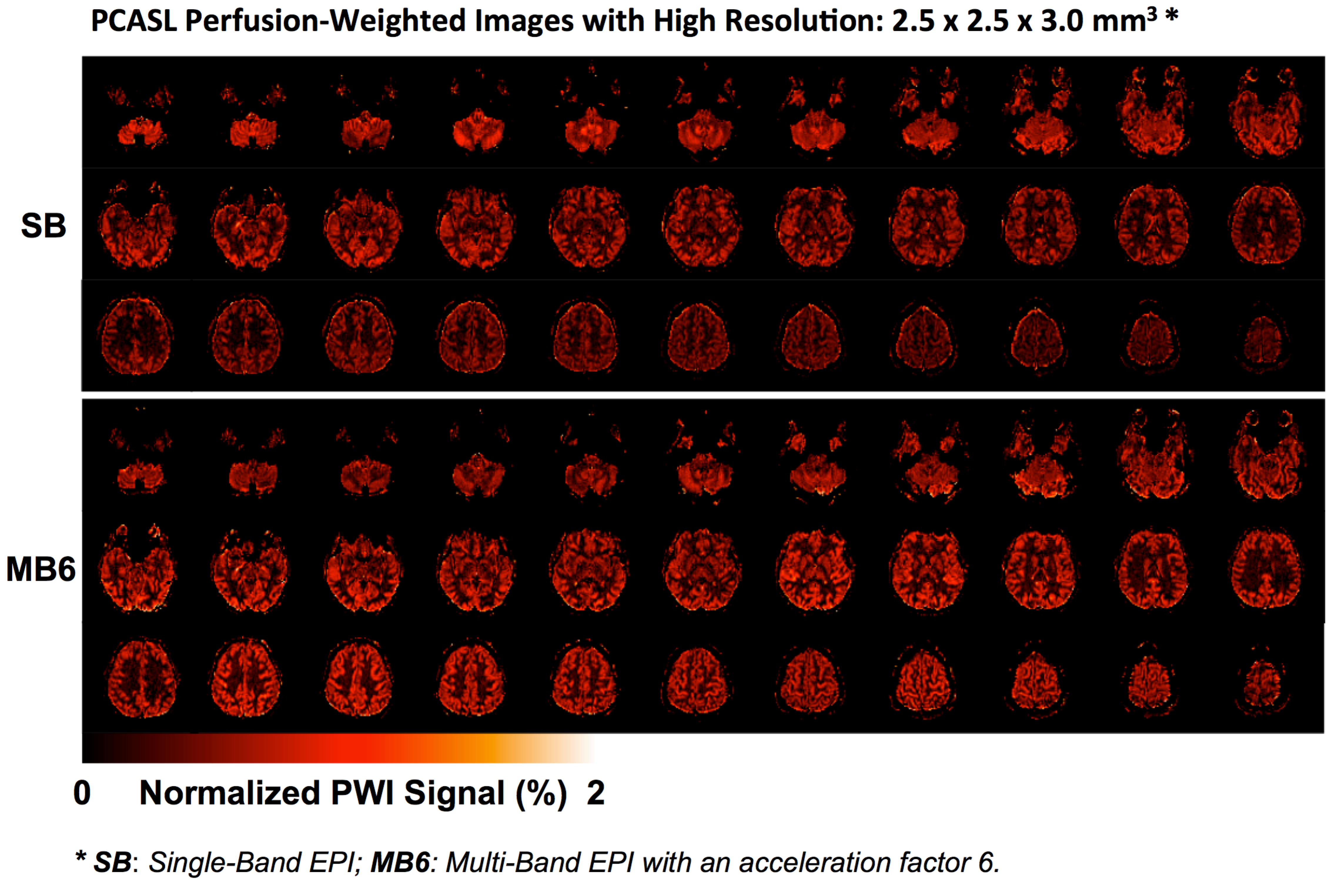
High-resolution arterial spin labeling (ASL) imaging is highly desirable in both neuroscience research and clinical applications to reduce partial volume effects on gray matter (GM) and white matter (WM) cerebral blood flow (CBF) quantification, to improve the ability to assess perfusion abnormality in sub-cortical brain structures, e.g. the hippocampus, and to identify small brain lesions. Decreased perfusion SNR for high-resolution imaging is a consequence of multiple factors including; 1) increased in-plane resolution, 2) increased through-plane resolution and 3) the need for more slices to cover the same volume resulting in prolonged delay times between labeling and signal acquisition during which labeled spins experience longitudinal relaxation. The necessity of increasing the number of label/control image pairs for sufficient perfusion SNR greatly increases the total imaging acquisition time, limiting the practice of acquiring high-resolution whole brain ASL perfusion data using traditional 2D echo planar imaging method (or called single-band (SB) EPI). To overcome such challenges, different strategies have been previously proposed to increase perfusion SNR of ASL methods: using pulsed- or pseudo-continuous arterial spin labeling (PCASL) coil and 3D imaging acquisition.
Multi-band imaging, or simultaneous multi-slice imaging, provides an attractive and alternative solution to reduce the total acquisition time of high-resolution whole brain imaging with 2D EPI, especially when increased spatial or temporal resolution is desired. Recently theoretical and experimental evaluation of MB-EPI for PCASL imaging [1] indicates that
- MB leakage contamination has minimal impact on CBF quantification in PCASL imaging
- MB improves SNR efficiency for high-resolution PCASL imaging using 2D EPI
- MB-EPI better supports the single-blood compartment model for CBF quantitation
In contrast to 3D imaging methods, MB-EPI does not suffer adverse within and across slice T2 blurring.
Features
- Some EPI parameters have been specifically customized to avoid errors in MB-EPI PCASL imaging protocol preparation or modification.
- The implemented MB-EPI PCASL sequence acquires two M0 images after label/control images series. The mean image of these two M0 images can be used for CBF quantification.
CMRR C2P
If you are interested in obtaining these sequences, the CMRR C2P instructions and agreement can be found on this page at the UMN Office for Technology Commercialization. Once you have executed a C2P agreement and have been given an access password, the sequence binaries can be downloaded here.
Note to users: If you have noticed a bug or have a request for a new feature in a future release, please email Xiufeng Li lixx1607@umn.edu to open a new support request. Be sure to include the sequence variant and the model of scanner you are using in the problem description.
Download the MB-EPI PCASL Sequence
|
MB-EPI PCASL Release 1.0 |
|
|
Released date MB-EPI PCASL Release Notes v1.0 |
Download
 Release 1.0 for VB17 (build) Release 1.0 for VB17 (build) |
References
1. Li X, Wang D, Auerbach EJ, Moeller S, Ugurbil K, et al. (2015) Theoretical and experimental evaluation of multi-band EPI for high-resolution whole brain PCASL Imaging. Neuroimage 106: 170-181.